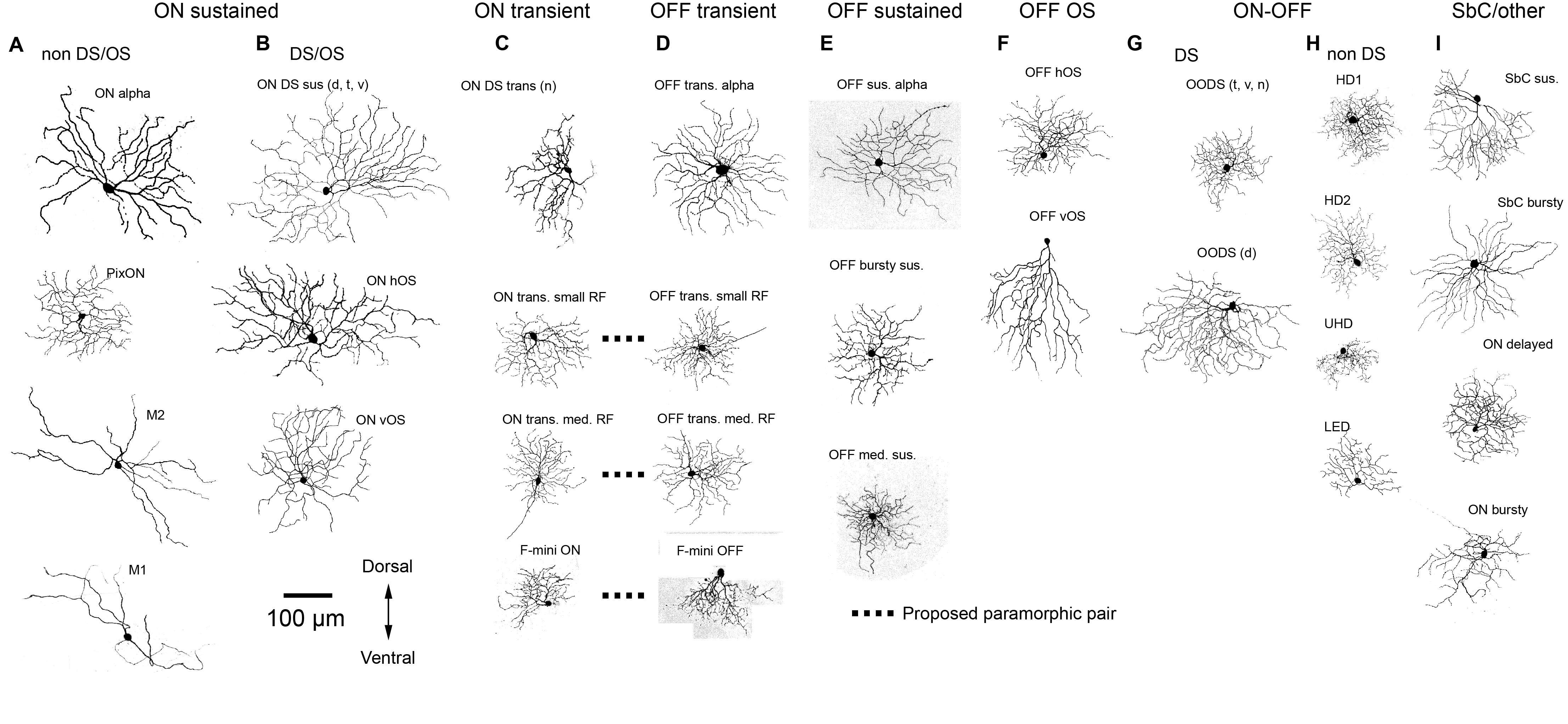
The retina is not a video camera; it is the world’s most advanced image processing machine, refined over hundreds of millions of years of evolution. My lab seeks to understand the computations performed by retinal circuits. We are working toward a comprehensive classification of retinal ganglion cells, the output cells of the retina, that includes physiology, morphology and gene expression patterns for each cell type. Our catalog of mouse retinal ganglion cells is online at rgctypes.org.
From this “parts list” we work our way into the circuits of the retina to answer the “how” questions about the synaptic and circuit mechanisms of retinal computation.
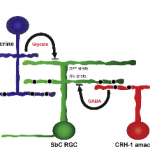
A circuit we discovered that involving two amacrine cell types and the sustained suppressed-by-contrast retinal ganglion cell.
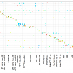
A heatmap showing genes (y-axis) that are selectively expressed in particular retinal ganglion cell types (x-axis).
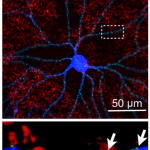
Identifying synaptic connections in the retina by triple-labeling the presynaptic (red) and postsynaptic (blue) cells and postsynaptic sites (green).
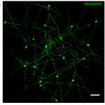
A network of amacrine cells coupled by gap junctions.
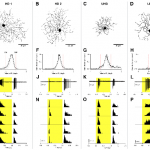
Four of the smallest, most abundant retinal ganglion cell types of the mouse, identified by morphology and physiology.
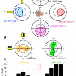
A subset of direction-selective and orientation-selective cell types of the mouse retina.
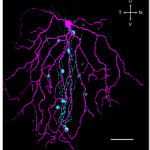
OFF orientation-selective retinal ganglion cells (magenta) inherit their feature selectivity from electrically coupled amacrine cells (cyan).